Anatomy 結構
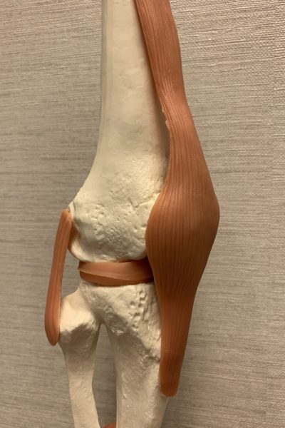
Knee is a joint made of 4 bones, Femur (the thigh bone), Patella (the knee cap), Tibia (the shin bone), Fibula (the smaller shin bone).
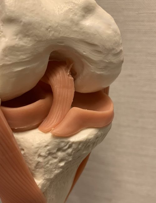
Two strong ligaments, Anterior Cruciate Ligament (ACL) and Posterior Cruciate Ligament (PCL), linked the femur and the tibia at the joint center, constraining abnormal translation front and back, as well rotation.
Another two ligaments, Medial Collateral Ligament (MCL) and Lateral Collateral Ligament (LCL), lie at inner and outer side of the knee , preventing the knee from opening up at side.
The knee cap travel at the valley of the femur, called Trochlea. A small ligament called Medial PatelloFemoral Ligament (MPFL) helps guiding its track. The tracking of the patella requires muscle coordination, congruent trochlea with sufficient depth, and an intact MPFL.
There are two meniscus inside the joint. Both are C shaped when view above, and triangular in cross section. Medial Meniscus locates at the inner and Lateral Meniscus is at the outer half of the knee. They make the curved articulating surface of femur having tight fit with the flat tibia surface. Without their prescence, it will be a point or line contact rather than a surface contact.


Pathology 毛病
Disease 病變
The followings are common pathologies in knee
以下是常見膝病變
- ACL tear 前十字韌帶撕裂
- Meniscal tear 半月板撕裂
- Degenerative knee 膝關節退化
- Osteochondral lesion 軟硬骨病變
- Patella dislocation 臏骨脫位
- MPFL tear 內側髕骨股骨韌帶撕裂
- Subchondral bone marrow edema 軟骨底骨髓水腫
- Varus knee 膝內曲
- Osgood-Schlatter disease 脛骨粗隆骨骺炎
Investigation 評估
MRI is usually required to review soft tissue status. Occasionally, standing X-ray would be needed.
磁力共震片能夠提供軟組織情況,所以很多時會是醫生的選擇。某些情況需安排站立受力X光。
Management 治療
Conservative treatments are indicated for majority of cases. They included,
- Physiotherapy
- Chiropractor
- TCM practitioner
- Prescribed Exercise
- Medication
For cases failed conservative treatments, or when the injury is unstable, then surgical intervention would be indicated.
For patients with witnessed deterioration, surgical intervention might be advocated.


絕大多數病人只需採取保守治療。他們包括
- 物理治療
- 脊醫
- 中醫
- 處方運動
- 藥物(口服及外敷)
對於保守治療無效的病例,或受傷位置不穩定,醫生會改為介入治療,例如打針甚至手術。
對於持續惡化的患者,醫生往往會建議手術,以保肢體功能。
Common Interventions include
- Injection of steroid
- Injection of PRP
- Injection of hyaluronic acid
- casting
- Arthroscopic lateral retinacular Release
- Arthroscopic chondroplasty and microfracture
- Arthroscopic meniscal repair
- Arthroscopic partial menisectomy
- ACL reconstruction
- Subchondroplasty
- Surgical reduction and plating
- Unicondylar Knee Replacement (UniKR)
- Total Knee Replacement (TKR)

